Peptide Price Finder

Home · Research · Other

GDF-8 (myostatin) research

What published studies found for GDF-8 (myostatin): 6 papers, 0 in humans, with sample sizes and key results.

C
Mostly animal studies
Why: No human studies give myostatin itself; mouse studies that administer it show muscle and fat loss and impaired healing. Antibody trials block it, a different treatment.

No human study has given GDF-8 (myostatin) itself as a treatment. The animal and cell work that does administer it shows harm or no benefit: in mice it caused cachexia-like muscle and fat loss and impaired bone and muscle healing, and it did not reduce restenosis. The human trials in this area test antibodies that block myostatin, a different treatment, so they are not evidence for products sold as "GDF-8".

✗ 3 negative? 3 inconclusive

No human safety data exist for GDF-8 given as a drug; in mice it caused cachexia-like wasting.

US compounding: Not compoundable. Not an FDA-approved drug and not nominated for FDA's 503A bulks list, so US pharmacies can't legally compound it. Status for every peptide.

Browse and filter these papers

Animal & lab studies

✗ NegativeanimalScience · 2002

Induction of cachexia in mice by systemically administered myostatin.

AnimalsSystemic overexpression of myostatin

Raising circulating myostatin in adult mice (via implanted myostatin-secreting cells) caused profound muscle and fat loss resembling cachexia.

Studied
Adult mice
Main outcome
Muscle and fat mass
Authors' conclusion
Systemic myostatin overexpression in adult mice induced profound muscle and fat loss analogous to human cachexia; the authors suggest blocking myostatin as a therapeutic target.
Limitations
Animal study; myostatin raised by overexpression rather than a measured dose.
✗ NegativeanimalJ Histochem Cytochem · 2012

Immunolocalization of myostatin (GDF-8) following musculoskeletal injury and the effects of exogenous myostatin on muscle and bone healing.

AnimalsAnimal injury model with hydrogel delivery of exogenous recombinant myostatin vs vehicle

In mice with deep musculoskeletal injury, hydrogel-delivered recombinant myostatin reduced fracture callus cartilage area (by 41% and 80% at two doses) and bone volume (by 30.6% and 38.8%) and increased fibrous tissue in muscle, i.e. it impaired healing.

Key resultCallus cartilage area fell 41% and 80% (10 and 100 µg/ml; p<0.05); callus bone volume fell 30.6% and 38.8% (p<0.05)
Studied
Mice with deep penetrant musculoskeletal injury (muscle and bone)
Compared with
Vehicle
Main outcome
Callus cartilage area and bone volume; fibrous tissue in muscle
Authors' conclusion
Exogenous myostatin worsened bone and muscle healing, suggesting early myostatin inhibition may improve regeneration after such injury.
Limitations
Mouse study with local hydrogel delivery; sample size not given in the abstract.
✗ NegativeanimalInt J Mol Sci · 2020

Myostatin Inhibits Vascular Smooth Muscle Cell Proliferation and Local 14q32 microRNA Expression, But Not Systemic Inflammation or Restenosis.

Humans and animalsIn vitro dose-response plus in vivo myostatin infusion

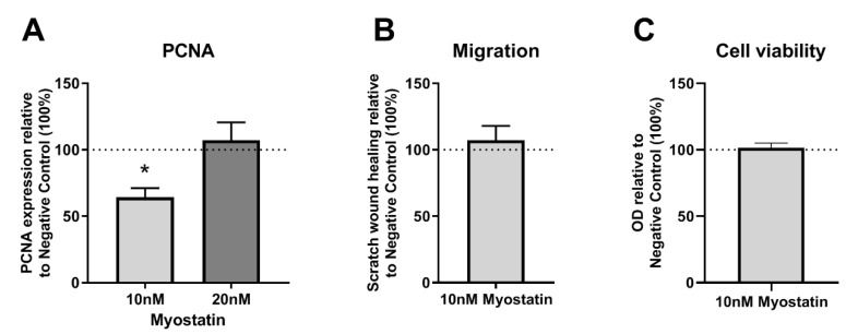
Myostatin reduced vascular smooth muscle cell proliferation and 14q32 microRNA expression in vitro, but infusing myostatin in mice did not reduce restenosis, neointimal area or lumen stenosis.

Studied
Cultured vascular smooth muscle cells and a mouse model of post-interventional restenosis
Main outcome
Restenosis, neointima, VSMC proliferation, 14q32 microRNA expression
Authors' conclusion
Myostatin did not affect post-interventional restenosis, though it inhibited VSMC proliferation and 14q32 microRNAs; it had no effect on macrophage activation or influx.
Limitations
Mouse model and cell culture; no human data.
Figure 3: (A–C): PCNA expression, cell migration and cell viability in myostatin treated VSMCs relative to non-myostatin treated control (100%). (A)—P
Figure 3. From PMC7278907, CC BY (terms); resized.
? Inconclusivein vitroBiomolecules · 2020

Differentiation of Murine C2C12 Myoblasts Strongly Reduces the Effects of Myostatin on Intracellular Signaling.

Cells / labIn vitro administration of myostatin with analysis of canonical and noncanonical signalling

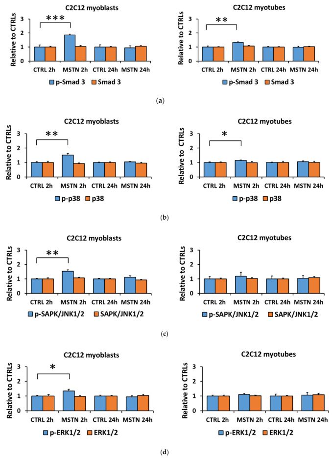
Myostatin applied to mouse C2C12 and human CHQ myoblasts and myotubes strongly signalled in myoblasts, but differentiated C2C12 myotubes largely lost the response (possibly via increased follistatin), while human CHQ cells stayed responsive.

Studied
Murine C2C12 and human CHQ myoblasts and myotubes
Compared with
Untreated cells; C2C12 with C26 colon cancer cell coculture
Main outcome
Intracellular signalling downstream of myostatin
Authors' conclusion
The effects of myostatin on signalling may be cell line- or organism-specific, and C2C12 myotubes seem a nonoptimal model for studying them.
Limitations
Cell culture methods study; no in vivo or clinical outcome.
Figure 1: Myostatin-induced changes in the canonical (Smad3) and noncanonical (MAPKs) pathways were greater in C2C12 myoblasts than they were in myotu
Figure 1. From PMC7277184, CC BY (terms); resized.
? Inconclusivein vitroEndocrinology · 2014

Myostatin stimulates, not inihibits, C2C12 myoblast proliferation.

Cells / labIn vitro proliferation assays with recombinant myostatin from two eukaryotic sources, TGF-beta1, and Alk4/5 inhibitors

Two eukaryote-made recombinant myostatin preparations stimulated, rather than inhibited, C2C12 myoblast proliferation (via Smad2), questioning earlier work with bacteria-made myostatin.

Studied
Mouse C2C12 myoblast cell line
Compared with
Serum, TGF-beta1; receptor inhibitors SB431542 and SB505142
Main outcome
Myoblast proliferation and Smad2/3 phosphorylation
Authors' conclusion
Myostatin and TGF-beta1 stimulate C2C12 proliferation mainly via Smad2, which the authors say questions the physiological relevance of the C2C12 model.
Limitations
Immortalised cell line only.
? Inconclusivein vitroPflugers Arch · 2021

Myostatin regulates the production of fibroblast growth factor 23 (FGF23) in UMR106 osteoblast-like cells.

Cells / labIn vitro myostatin treatment with pathway inhibitors; qRT-PCR and ELISA

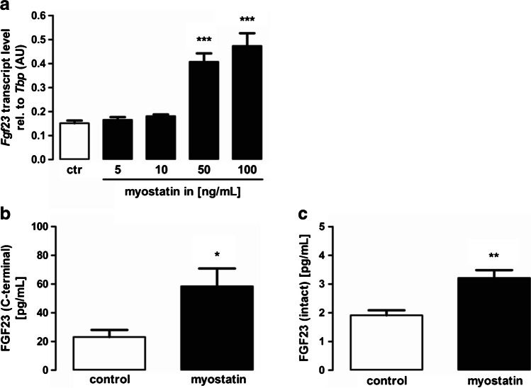
Myostatin raised Fgf23 gene expression and FGF23 protein production in UMR106 osteoblast-like cells, partly through TGF-betaRI/p38MAPK and NFkB-dependent calcium entry; a signalling study in a rat cell line.

Studied
UMR106 osteoblast-like cells
Compared with
Untreated cells; inhibitors of TGF-betaRI, p38MAPK, NFkB and SOCE
Main outcome
Fgf23 gene expression and FGF23 protein
Authors' conclusion
Myostatin stimulates Fgf23 expression in these cells, an effect at least partly mediated by TGF-betaRI/p38MAPK signalling and NFkB-dependent store-operated calcium entry.
Limitations
Single cell line; no in vivo or human data.
Fig. 2: Myostatin induces Fgf23 expression in UMR106 cells, a Arithmetic means ± SEM of relative (rel.) Fgf23 mRNA abundance or b C-terminal or c in
Fig. 2. From PMC8164604, CC BY (terms); resized.

Verdicts are judged on each paper's own main outcome; grades run from A (strong human evidence) to D (little or none). Details are read from the abstracts, and figures are reproduced only from openly licensed full texts. Not medical advice.